
Techniques
Ultrafast Electron techniques for microscopy, crystallography and spectroscopy

Ultrafast processes - events that unfold on femtosecond timescales - remained invisible for decades. These processes occur too fast to be captured by conventional instruments, leaving entire classes of physical, chemical, and biological phenomena effectively out of reach. Our technology changes that.
Ultrafast Crystallography
Study structural dynamics at atomic length and timescales using ultrafast electron diffraction (UED). Capture how crystal structures change during phase transitions, chemical reactions, and rapid processes.

Ultrafast Microscopy
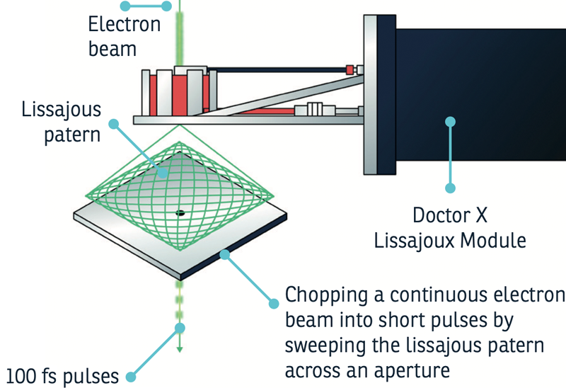
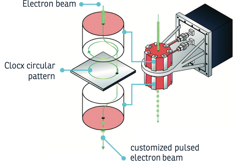
The Lissajoux Module and Clocx Module, both compatible with XFEG and CFEG systems, add ultrafast capability to your existing transmission electron microscope without compromising performance.

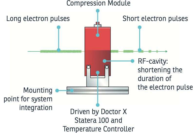
Single shot femtosecond UED
van Oudheusden et al., Phys. Rev. Lett. 105, 264801 (2010)
In 2010, the Eindhoven University of Technology group demonstrated what many thought impractical: compressing space-charge-dominated electron bunches to sub-100 fs durations while retaining enough charge for single-shot diffraction.
The challenge: to capture a diffraction pattern in one shot, you need roughly a million electrons (0.1–0.2 pC) in a single bunch. But Coulomb repulsion rapidly stretches any dense electron bunch - femtoseconds become picoseconds within centimeters of drift. Previous approaches either accepted longer pulses or used multiple shots, limiting the choice of samples and experimental repeatability.
The solution: an RF cavity operating at 3 GHz in TM-010 mode, synchronized to the photoemission laser. The oscillating longitudinal field decelerates electrons at the front of the bunch and accelerates those at the back, inverting the space-charge-induced velocity spread. As the bunch drifts downstream, it compresses—100-fold, from 10 ps back to sub-100 fs.
The group demonstrated this with a transmission diffraction experiment on polycrystalline gold foil. A single 200 fC bunch at 95 keV produced a high-quality diffraction pattern with clearly resolved Debye-Scherrer rings. Arrival time jitter: 80 fs.
This work established RF compression as the enabling technique for single-shot femtosecond electron diffraction—the foundation for Doctor X’s Structural Dynamics Probe.
Phase transitions in VO2
Morrison et al., Science 346, 445–448 (2014)
Vanadium dioxide (VO2) switches from semiconductor to metal at around 70°C—a transition fast enough to interest device engineers, but poorly understood. Does the electronic structure drive the change, or does the lattice? The two have always moved together, making them impossible to separate with conventional techniques.
Morrison et al. used ultrafast electron diffraction combined with infrared transmissivity to pull them apart.
By photoexciting the monoclinic semiconducting phase with carefully tuned laser pulses, they induced a transition to a metastable state that had never been observed before: the material acquired metal-like mid-infrared optical properties while retaining the periodic lattice distortion characteristic of the semiconductor. The lattice stayed put; the electrons reorganized anyway.
This decoupling—lattice frozen, electrons mobile—demonstrated that the electronic transition can proceed independently of the structural one. It also showed that ultrafast electron diffraction can follow both lattice and electronic dynamics on the same timescale, providing complementary information to X-ray techniques.
The experiment exemplifies what ultrafast crystallography enables: separating entangled degrees of freedom by catching them at different moments in time.
Advancing ultrafast (S)TEM
Struct. Dyn. 12, 064303 (2025)
Transmission electron microscopy (TEM) has significantly advanced fields such as materials science, nanotechnology, and structural biology by providing detailed structural and analytical information at picometer resolutions. To further enhance TEM’s capabilities, time-resolved electron microscopy introduces the temporal domain, using ultrafast electron pulses to capture dynamic processes.
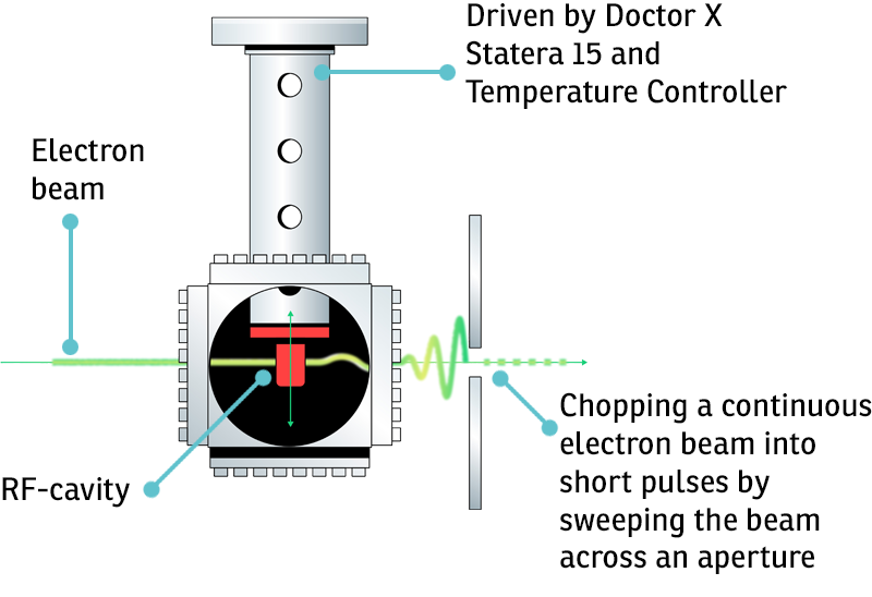
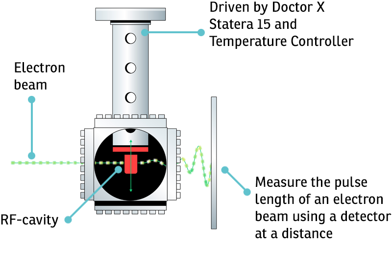
Traditional methods generate these pulses via photocathode illumination by femtosecond lasers and face challenges like complex alignment and limited repetition rates. An alternative approach employing electronic devices as beam choppers, specifically resonant RF cavities in combination with electrostatic beam blankers, simplifies alignment and increases repetition rates, achieving picosecond and sub-picosecond pulses.
Additionally, these devices do not compromise the performance of the microscope in any other imaging mode. The microscope can be rapidly toggled between continuous and pulsed imaging, providing flexibility of operation in modern research labs. This study integrates these beam choppers into high-end TEMs and demonstrates their effectiveness in achieving high temporal resolution for pump–probe experiments.
Results show that these methods maintain high spatial resolution and coherence, making them a promising solution for ultrafast electron microscopy

Prof. Dr. Ir. Jom Luiten
Concepts Into Products
The technology originated from research at Eindhoven University of Technology, where Jom Luiten developed fundamental ultrafast electron techniques using ultrafast electron guns and Radio Frequency cavities to precisely control the electron pulses. Turning these into reliable products required rethinking how components work together across physics, electronics, mechanics, and software, then standardizing everything so it could be manufactured repeatably and operate in real lab conditions.
Every instrument is built for durability. Most of the development focuses on turning concepts into reliable products that can survive years of daily use. Every design decision assumes someone will depend on this equipment late at night, under deadline pressure, when funding and research timelines are at stake.
Our DNA
Doctor X was founded in 2018 by brothers Jom and Thomas Luiten. Jom spent years at Eindhoven University of Technology developing ultrafast electron techniques - building instruments that didn’t exist yet. With results came demand for his custom-built instruments.

Doctor X makes research instruments that give scientists high time and energy resolution at a precision that wasn’t accessible before.

X LAB is where Doctor X develops the next generation of ultrafast electron techniques. This is the heart of the company - where ideas become instruments.
Research here pushes beyond current products. Rotating mode cavities for faster pulse repetition. Pulse shaping for application-specific beam profiles. X-ray generation through the Smartlight project. Each direction opens new capabilities for researchers who need them.
The work happens the same way it always has at Doctor X: making, testing, refining, and sometimes discarding. Not every idea becomes a product. The ones that do will extend what ultrafast electron techniques can unlock - and what researchers can discover.